Anti-mouse CD19 nanobeads bind CD19⁺ B cells directly and are retained on a column in a magnetic field. In mouse spleen the manual reports 55.6% CD19⁺ before separation and 94.4% after a single positive selection, with 1.54% residual CD19⁺ cells in the flow-through. CD19 is expressed from pro-B to mature B cells and on follicular dendritic cells; plasma cells are negative.
| Catalogue | Size | List price | Promo price | Manual | COA |
|---|---|---|---|---|---|
| PM019L | 2 mL, for 2e+9 cells | ¥4000 | ¥3200 | Search | |
| PM019M | 1 mL, for 1e+9 cells | ¥2500 | ¥2000 | Search | |
| PM019S | 0.2 mL, for 2e+8 cells | ¥700 | ¥560 | Search |
Validation data
Mouse spleen was labelled with anti-mouse CD19 Nanobeads at 10 μL per 1×10⁷ cells, incubated at 4 °C for 10 min and separated on a column. The eluted positive fraction and the flow-through were stained with APC anti-mouse CD19 and analysed by flow cytometry.
| Readout | Result |
|---|---|
| CD19⁺ before separation (mouse spleen) | 55.6% |
| CD19⁺ purity after one positive selection (positive fraction) | 94.4% |
| CD19⁺ carry-over in the flow-through (depleted fraction) | 1.54% |
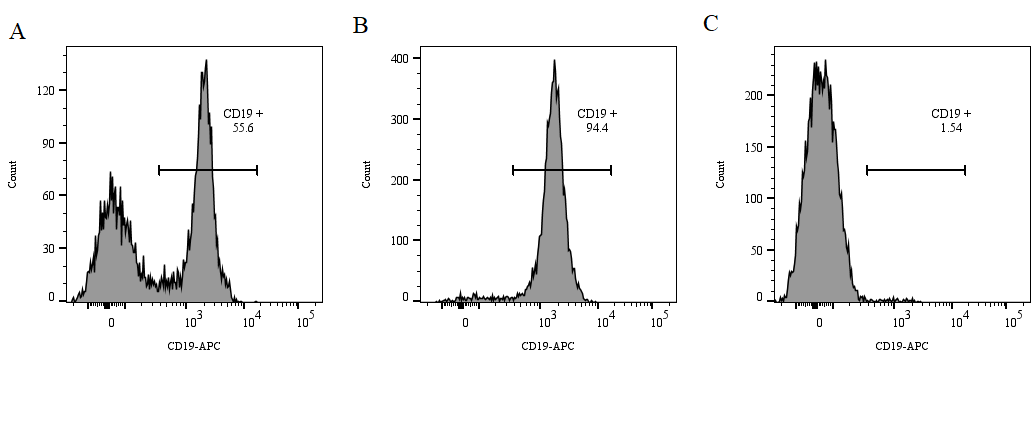
94.4% refers to CD19⁺ cell purity — predominantly B cells, with a possible minor FDC component; plasma cells are CD19-negative and outside the selected fraction. Data come from a single sample and a single separation without absolute cell counts, so they are not converted to yield. Tissues with a lower starting frequency, such as bone marrow, give lower purity on a single column.
Frequently asked questions
Does CD19 positive selection yield a pure B-cell population?
CD19 is expressed from pro-B through mature B cells and on follicular dendritic cells (FDCs); plasma cells are negative. The selected fraction is therefore a CD19⁺ population that is predominantly B cells but may contain a small number of FDCs. To resolve transitional, follicular or marginal-zone subsets, add CD23, CD43, IgD or IgM to the panel.
Are plasma cells present in the selected fraction?
No. Plasma cells downregulate CD19 after terminal differentiation, so they fall outside the CD19⁺ gate. If antibody-secreting cells are the target, use a plasma-cell marker such as CD138 or run ELISPOT on the immunised tissue directly.
How is mouse spleen prepared, and what starting frequency should I expect?
Mince the spleen, filter through a 70 μm sterile nylon mesh, lyse red blood cells and count (AO/PI staining recommended). CD19⁺ cells typically account for 10–60% of mouse spleen (55.6% in the manual validation sample); one spleen yields roughly 0.5–1×10⁸ cells.
How much bead is needed, and does the dose vary with starting frequency?
At 1×10⁸ cells/mL, dose by CD19⁺ frequency: 1–5 μL per 1×10⁷ cells for tissue samples below 10%, and 10 μL per 1×10⁷ cells for mouse spleen at 10–60%. Keep labelling at 4 °C for 10 min; do not increase the amount to chase purity, as excess bead raises non-specific binding.
Which column and magnet combination should I use?
M column: <1×10⁷ labelled cells / <2×10⁸ total cells, with magnet M1/M8, 500 μL equilibration and 3×500 μL washes. L column: <1×10⁸ labelled cells / <2×10⁹ total cells, with magnet L1/L4, 3 mL equilibration and 3×3 mL washes. One mouse spleen (0.5–1×10⁸ cells) normally fits an M column; size by total cell count, not by the post-filter estimate.
Can the same kit deplete CD19⁺ cells instead?
Yes. The flow-through from loading and washing is the CD19-depleted fraction; the manual reports 1.54% residual CD19⁺ cells. This suits non-B-cell enrichment or control arms in B-cell-dependent functional assays, with no second reagent required.
Can I run a second column if purity is too low?
Yes, but judge whether it is worth it. When CD19⁺ frequency is low (bone marrow, blood), repeat the equilibrate–load–wash–elute cycle on a fresh column to gain purity. At normal spleen frequency (55.6%) a second pass mainly costs cells and is usually unnecessary.
Why FACS buffer rather than PBS?
PBS lacks protein protection and osmotic buffering, which lowers recovery and viability. Use FACS buffer (1× PBS with 0.02% EDTA and 2% FBS) or Eynos separation buffer; buffer quality often matters more than the bead itself.
Should flow-cytometry antibody amounts be adjusted after sorting?
Yes. At 94.4% purity the target cells are far more concentrated per unit volume than in the starting sample, so add antibody according to total cell number rather than reusing the pre-sort volume; otherwise under-staining will distort intensity and gating.
Can bone marrow also reach 94.4% purity?
Not directly. The 94.4% figure comes from mouse spleen (55.6% starting). CD19⁺ frequency in bone marrow is much lower, so a single column gives lower purity and a second pass is usually needed. Starting frequency and subset composition differ between tissues and mouse strains, so each source should be benchmarked.
How do I avoid air bubbles when eluting cells from a column?
Bubbles usually come from protein-containing buffer. Fill the column with 3 mL of FACS buffer before pushing the cells out, and press the plunger once — do not pump repeatedly. If bubbles persist, switch to a high-quality PBS for the elution step.